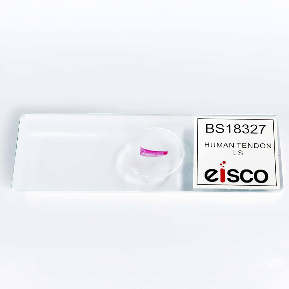
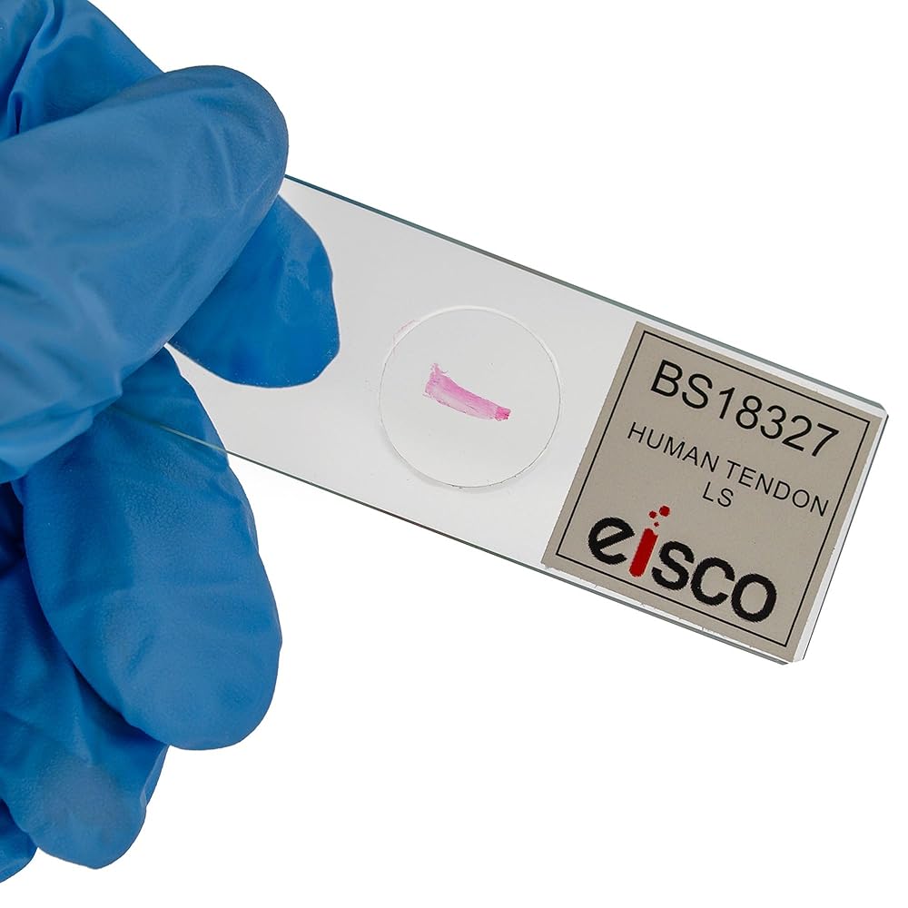
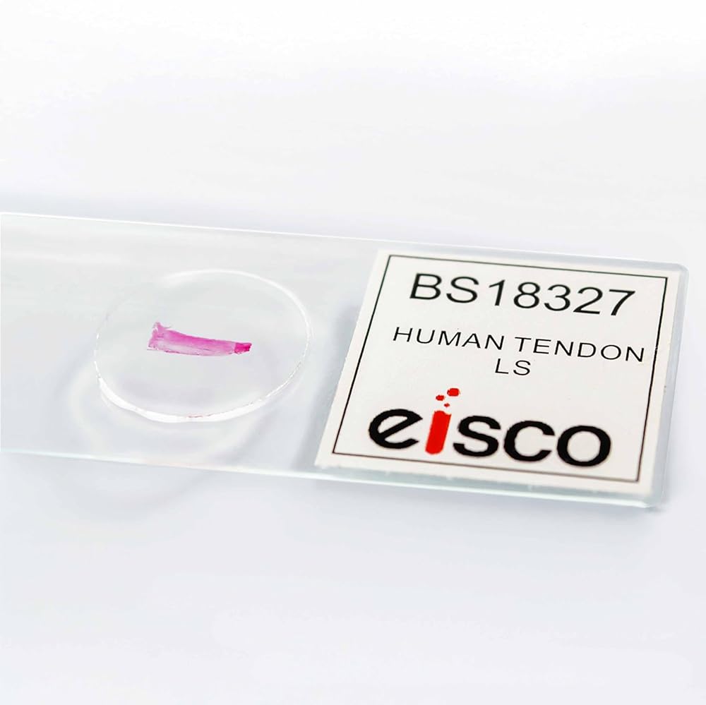
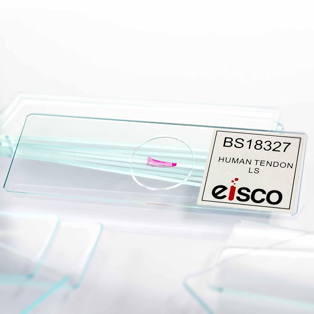
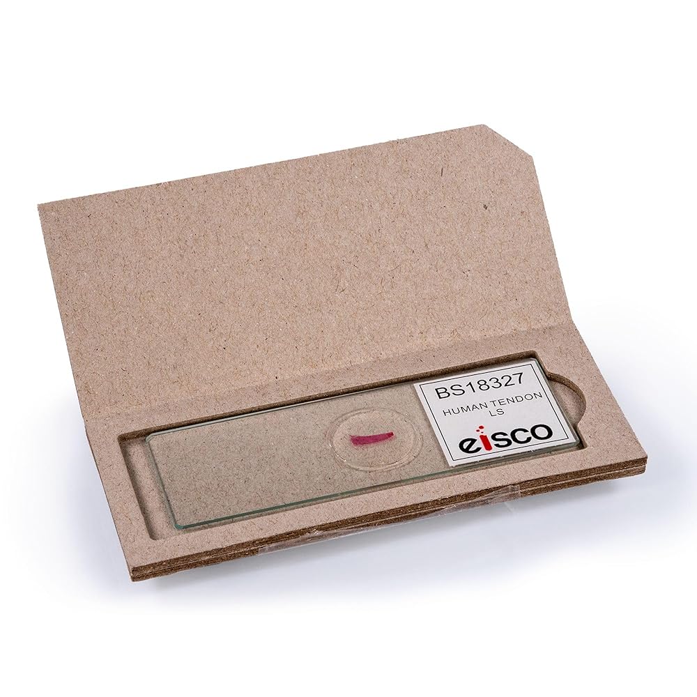
HUMAN TENDON PREPARED SLIDE || Includes a longitudinal section (LS) of a human tendon tissue mounted on a labeled glass slide; For microscopic observation of structural elements
VISIBLE FIBER ALIGNMENT || Clearly displays the parallel arrangement of collagen fibers associated with connective tissue; Supports identification of tendon composition and orientation
STRUCTURAL INSIGHT || Allows learners to visualize tendon structure, relating to muscle attachment and mechanical function, reinforcing understanding of musculoskeletal connective tissues
SUITABLE FOR TEACHING AND STUDY || Useful in biology, anatomy, and histology instruction; Ideal for lab activities and hands-on study in classroom, university or clinical settings
GLASS SLIDE CONSTRUCTION || Mounted on a standard 75 x 25 mm glass slide with sealed coverslip; Durable design for repeated use with standard compound microscope stages
وصف
This prepared microscope slide features a longitudinal section of human tendon tissue mounted and sealed for microscopic examination. Clearly reveals the parallel alignment of collagen fibers and overall tendon structure. Supports instruction in histology, anatomy, and musculoskeletal function. Ideal for classroom demonstrations, lab-based learning, and anatomical tissue identification using standard compound microscopes.
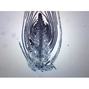 طرف جذع Elodea - مقطع عرضي - شريحة مجهرية مُجهزة - 75 × 25 مم - علم الأحياء والمجهر - Eisco Labs
KWD 3
طرف جذع Elodea - مقطع عرضي - شريحة مجهرية مُجهزة - 75 × 25 مم - علم الأحياء والمجهر - Eisco Labs
KWD 3
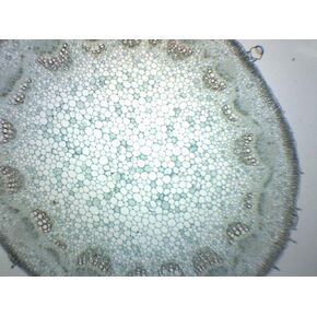 Helianthus Stem - شريحة مجهرية مُجهزة - 75 × 25 مم - علم الأحياء والمجهر - Eisco Labs
KWD 3
Helianthus Stem - شريحة مجهرية مُجهزة - 75 × 25 مم - علم الأحياء والمجهر - Eisco Labs
KWD 3
 مستورقات، حامل كامل - شريحة مجهرية مُجهزة - 75 × 25 مم - علم الأحياء والفحص المجهري - Eisco Labs
KWD 4
مستورقات، حامل كامل - شريحة مجهرية مُجهزة - 75 × 25 مم - علم الأحياء والفحص المجهري - Eisco Labs
KWD 4
 مفتاح الاتصال Eisco Labs، رمز تيليغرافينغ/مورس، مفرد
KWD 5.500
مفتاح الاتصال Eisco Labs، رمز تيليغرافينغ/مورس، مفرد
KWD 5.500